Colorectal cancer usually begins as a small, seemingly harmless cluster of cells called a polyp. Finding the precise, jagged edge of that polyp on a colonoscopy screen is the difference between catching a fatal threat early and sending a patient home with a false sense of security.
For years, computer scientists have trained artificial intelligence to hunt for these lesions using a basic mathematical strategy: addition. To find a tumor, an AI gathers the big picture context of an organ and adds it to the microscopic details of the tissue. But what if this instinct to accumulate data is exactly what makes the AI blind to the most critical borders?
The Problem with Addition
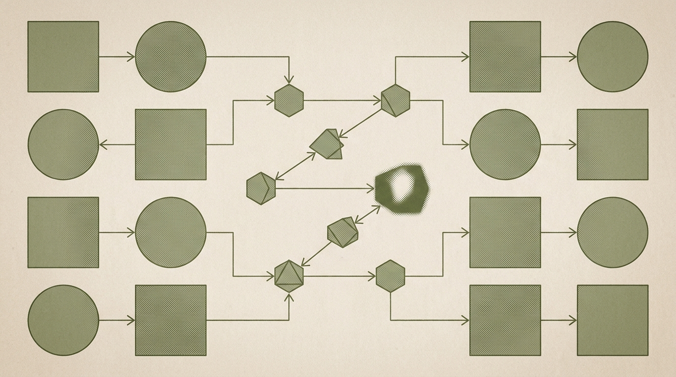
When an algorithm looks at a medical scan, it generates abstract grids of numbers called feature maps. To accurately draw a boundary around a newly formed lung infection or a breast tumor, the algorithm must merge maps that contain zoomed-out semantic context with maps full of zoomed-in textures. Traditionally, developers combined these views using element-wise addition or by stacking the layers side-by-side.
This habit creates a massive amount of data redundancy. Blending two highly correlated layers of visual information does not yield a sharper image. It behaves more like a loud, redundant echo that washes out subtle transitions in the tissue. The more information the AI stacks together, the blurrier the boundaries become.
So, how can an algorithm filter out the noise to find the true edge of a lesion?
Noise-Canceling Mathematics
The answer involves completely reversing the math. A new network architecture, named M2SNet, replaces addition with subtraction.
When you subtract a broad feature map from a highly detailed one, the shared, redundant information mathematically cancels out. What remains is a pure, highly refined map of the differences. It functions exactly like noise-canceling headphones, silencing the duplicated background echo to amplify the structural edges of the tumor.
This subtraction takes place inside specialized mathematical blocks throughout the network. However, medical anomalies come in wildly different shapes and sizes. An algorithm optimized for a large mass might completely overlook a tiny cyst. To fix this, researchers had to teach their subtraction blocks to simultaneously recognize varying structures.
But how do you train an AI to process multiple scales without bogging it down with millions of new, complex calculations?
Grading for Comprehension
Instead of relying on heavy, parameter-bloated modules, the network uses simple mathematical filters filled entirely with the number one. Multiplying the data by these fixed grids naturally captures distinct regional clusters, from isolated pixels to large textures, without increasing the computational burden on the system.
Processing the image is only half the battle. The AI also has to be graded on its accuracy so it can learn from its mistakes. Traditionally, researchers grade an algorithm by comparing its prediction to a human doctor’s hand-drawn outline, strictly pixel by pixel.
To break this habit of rote memorization, researchers introduced a secondary grading system called LossNet. LossNet is a pre-trained image classifier. Even though it was originally trained on everyday photos of dogs and cars, it inherently understands universal visual rules about edges and textures. LossNet acts as an automated teacher by taking the AI’s prediction and the true answer, and comparing their underlying structural logic. It is similar to grading a student’s essay based on their thesis and logical flow, rather than simply docking points for minor spelling errors.
The Subtraction Payoff
By combining mathematical subtraction with this intuitive grading system, the algorithm dramatically outperforms conventional models.
When tested globally across four wildly different medical imaging tasks, colon polyps, ultrasound breast tumors, CT lung infections, and optical eye layers, it consistently drew sharper, more accurate boundaries. More impressively, it achieved this using only a fraction of the computational power of its competitors, operating seamlessly at a blazing 90 frames per second.
The hunt for microscopic polyps and tumors proves a counterintuitive reality about computer vision. In a field obsessed with accumulating the largest possible datasets and stacking thick layers of deep neural networks, clarity does not always come from gathering more information. Sometimes, the only way to see a threat clearly is to strip away the data you already have and look closely at what gets left behind.